Introduction to Tendon Physiology and Peptide Research
Tendon injuries represent a significant challenge in orthopaedic research due to the tissue's intrinsic physiological limitations. Unlike muscle or skin, tendons possess a relatively poor vascular supply and a low metabolic rate, factors that contribute to slow repair processes and a high propensity for scar tissue formation rather than functional regeneration. Consequently, the scientific community has increasingly turned its attention to biological agents that may modulate the phases of tissue healing.
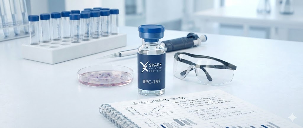
Among the peptides currently under investigation, BPC-157 (Body Protection Compound-157) has emerged as a primary subject of interest. A stable pentadecapeptide originally isolated from human gastric juice, BPC-157 has been studied extensively in preclinical models for its potential influence on soft tissue repair. Current research focuses on understanding how this peptide interacts with cellular pathways involved in angiogenesis, collagen synthesis, and inflammatory modulation within the context of tendon pathophysiology.
The Mechanism of Action: Angiogenesis and VEGF
One of the most cited mechanisms in the literature regarding BPC-157 is its potential interaction with the angiogenic process. Healing in hypovascular tissues like tendons is often rate-limited by the delivery of oxygen and nutrients.
Upregulation of Vascular Endothelial Growth Factor (VEGF)
Research utilizing cellular and animal models suggests that BPC-157 may influence the expression of Vascular Endothelial Growth Factor (VEGF). VEGF is a critical signaling protein that stimulates the formation of new blood vessels. In preclinical studies involving tendon transection, subjects treated with BPC-157 have demonstrated an increase in vascular density at the site of injury. This 'angiogenic effect' is hypothesized to be a core driver of the peptide’s regenerative potential, theoretically allowing for improved metabolic support during the proliferative phase of healing.
Modulation of the Nitric Oxide (NO) System
Scientific inquiry has also linked BPC-157 to the nitric oxide (NO) system. The NO pathway is complex; it regulates vasodilation and endothelial cell function. Studies propose that BPC-157 may act as a stabilizer of the NO system, potentially protecting endothelial tissue and facilitating the survival of new vessels formed during the repair process. This interaction suggests a multifaceted approach to revascularization that extends beyond simple growth factor stimulation.
Related Research: See GHK-Cu and Tissue Regeneration data.
Fibroblast Proliferation and Collagen Organization
The structural integrity of a tendon is defined by its extracellular matrix, which is primarily composed of Type I collagen. The cells responsible for synthesizing this matrix are tenocytes (tendon fibroblasts).
Stimulating Cellular Migration
In vitro studies on tendon fibroblasts have observed that exposure to BPC-157 may accelerate the rate of cell proliferation and migration. For a tendon defect to close, fibroblasts must migrate to the site of injury and proliferate in sufficient numbers to lay down a new matrix. Research data indicates that BPC-157 may enhance this cellular motility, potentially through the activation of the FAK-paxillin pathway, a signaling cascade involved in cell adhesion and migration.
Collagen Synthesis and Fiber Alignment
Quality of repair is as critical as the speed of repair. In many natural healing scenarios, the body produces disorganized Type III collagen (scar tissue), which is mechanically inferior to the original Type I collagen. Preclinical investigations have analyzed the histological quality of tendons following BPC-157 administration. Some findings suggest that the peptide may promote a more organized arrangement of collagen fibers, potentially influencing the ratio of collagen types produced during the remodeling phase.
Interaction with Growth Hormone Receptors
A more specific avenue of research involves the interaction between BPC-157 and the growth hormone (GH) axis within tendon tissue. Systemic growth hormone plays a vital role in tissue maintenance, but its local effects are mediated by receptors on the cell surface. Research utilizing tendon fibroblast cultures suggests that BPC-157 may increase the expression of growth hormone receptors on these cells. By potentially upregulating receptor density, the peptide could theoretically amplify the sensitivity of tendon cells to circulating growth factors.
Related Research: Review MOTS-c and Metabolic Signaling.
Preclinical Observations in Specific Tendon Models
The efficacy profile of BPC-157 has been mapped out through various animal models, typically involving rats or rabbits.
- Achilles Tendon Transection: In models involving complete or partial transection, BPC-157 administration often correlates with improved functional recovery metrics and higher load-to-failure ratios in biomechanical testing.
- Tendon-to-Bone Healing: The enthesis is notoriously difficult to heal. Preclinical data indicates BPC-157 may facilitate regeneration of the fibrocartilaginous zone at the tendon-bone interface.
- Muscle-Tendon Junction: Research has extended to the musculotendinous junction, investigating the ability to mitigate atrophy following contusion injuries.
Future Directions and Conclusion
While the existing body of literature regarding BPC-157 and tendon healing is robust within the preclinical sphere, it remains a subject of laboratory investigation. Future research aims to further elucidate the precise molecular signaling pathways involved, specifically distinguishing between direct effects on tenocytes versus indirect microenvironmental effects.
Conclusion: BPC-157 represents a significant focal point in the study of soft tissue regeneration. Through mechanisms involving angiogenesis, fibroblast modulation, and growth hormone receptor interaction, it offers a unique window into the molecular complexities of tissue repair.